Upper Leg Tendon Anatomy : Upper Thigh Muscles Diagram Quizlet - •medial thigh muscles•adductor longus muscle•adductor magnus muscle•adductor.. The vastus laterails works with the other quad muscles to help extend your knee joint. Upper leg tendon anatomy : Upper leg muscles and tendons : Anterior muscles extend your legs and flex your thighs. Anatomy_of_thigh_muscle 3/3 anatomy of thigh muscle anatomy of thigh muscle if you ally obsession such a referred anatomy of thigh muscle book that will allow you worth, get the categorically best seller from us currently from several preferred authors.
Muscles of the arm at la. The posterior upper leg muscles provide your knees with mobility (extension, flexion and rotation) and strength. The medial thigh muscles · adductor longus: The lower leg lies between the knee and ankle and works with the upper leg and foot to help perform the key functions of the leg. The thigh muscles don't just move your legs.
Tendinitis And Bursitis Treatment Cincinnati Tendinitis Dayton Oh from www.beaconortho.com Squeeze your knees together and boom, you're contracting the adductors. Anterior muscles extend your legs and flex your thighs. Tendons are cords made of tough tissue, and they work as special connector pieces between bone and muscle. The tendons for these muscles begin at your ischial tuberosity, or ischium (the bony bump under each buttock), and attach on the outer edges of your shinbones (tibia and fibula) just below the back of your knee. The hamstring portion of the adductor magnus has a similar action to these muscles, but is located in the medial thigh. The thigh muscles don't just move your legs. Its muscle belly is on the back aspect of the upper arm. It is also visible on the medial edge of the thigh from the anterior.
Ebraheim's educational animated video describes muscle anatomy of the thigh.
Like the biceps brachii in the arm, the biceps femoris muscle has two heads. Collectively, the muscles in this area plantarflex and invert the foot. / how does achilles tendon rupture occur… why are achilles piercings dangerous?. 3d anatomy tutorial on the muscles of the thigh and the gluteal region from anatomyzone for more videos, 3d models and notes visit: It's the area that runs from the hip to the knee in each leg. This is the group of muscles that you often see body builders flexing, which protrude just above the knee and take up most of the upper leg. If you feel it you need to take care of the causes of this hard pain. Upper leg muscle pain is a very hard pain affect the leg pain as a whole. The hamstring portion of the adductor magnus has a similar action to these muscles, but is located in the medial thigh. Upper leg anatomy and function the upper leg is often called the thigh. The medial, or towards the middle of the body, upper leg. It is the junction of the thigh and the leg and is a hinge joint. Meanwhile, the vastus lateralis is on the side of the thigh, while the vastus intermedius is hidden below the rectus femoris(5).
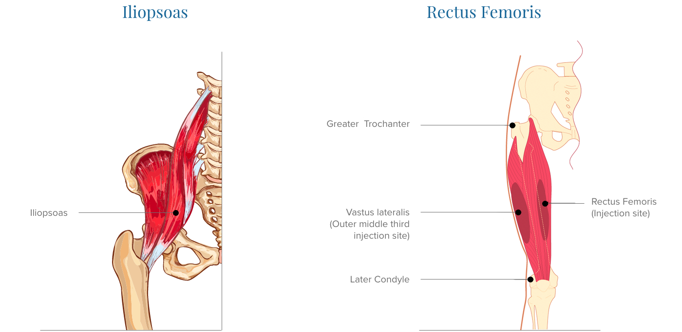
Quadriceps tendon attached superior and patellar ligament inferior to patella. The rectus femoris is located in the center of the thigh, while the vastus medialis is in the middle of the said body part. The patellar tendon is an essential tendon in the leg that enables the muscles to. The vastus laterails works with the other quad muscles to help extend your knee joint. It flexes the thigh at the hip joint, and extends at the knee joint.
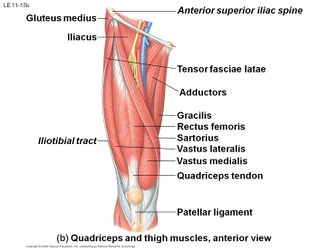
Quadriceps Tendon Tear Physiopedia from www.physio-pedia.com There are a number of bones, muscles, and tendons in the area. In clinical anatomy the thigh muscles are divided into three groups: The medial thigh muscles · adductor longus: Upper leg tendon anatomy : The posterior upper leg muscles provide your knees with mobility (extension, flexion and rotation) and strength. They consist of the rectus femoris, vastus intermedius, vastus lateralis and the vastus medialis. This is the group of muscles that you often see body builders flexing, which protrude just above the knee and take up most of the upper leg. If you want to droll books, lots of novels, tale, jokes, and more
Upper leg tendon anatomy :
Abdominal muscle diagram 12 photos of the abdominal muscle diagram abdominal muscle anatomy bodybuilding, abdominal muscle diagram female, abdominal muscle groups diagram, human abdominal muscle diagram, lower abdominal muscle diagram, human muscles, abdominal muscle anatomy bodybuilding, abdominal muscle diagram. It is also visible on the medial edge of the thigh from the anterior. The thigh has three sets of strong muscles: It's the area that runs from the hip to the knee in each leg. This tendon helps your leg bend when you raise your knee. The anterior, or front upper leg muscles are the quadriceps. They have a lot to do with how your hips move. It is the junction of the thigh and the leg and is a hinge joint. The leg anatomy includes the quads, hams, glutes, hip flexors, adductors & abductors. This is the group of muscles that you often see body builders flexing, which protrude just above the knee and take up most of the upper leg. The hamstring muscles in the back of the thigh, the quadriceps muscles in the front, and the adductor muscles on the inside. They consist of the rectus femoris, vastus intermedius, vastus lateralis and the vastus medialis. The quadriceps tendon attaches the quadriceps muscles to the patella.
The thigh has three sets of strong muscles: Upper leg muscle pain is a very hard pain affect the leg pain as a whole. The vastus laterails works with the other quad muscles to help extend your knee joint. Medial muscles adduct and rotate your thigh, and posterior flex your leg and extend your thigh. They have a lot to do with how your hips move.
Upper Leg Muscles Common Names Archives Anatomy Body Charts Muscle Anatomy Leg Anatomy Muscle Diagram from i.pinimg.com It flexes the thigh at the hip joint, and extends at the knee joint. They have a lot to do with how your hips move. This is why you have to indicate which biceps you are taking about when discussing one or other of these muscles. When we knowing the causes of the upper leg muscle pain it will be simple to treat and relieve the pain. Lateral (fibular) collateral ligament (fcl) upper part middle part lower part popliteus tendon (pt) upper part i. The posterior, or back, leg muscles for the upper leg are the hamstrings. The knee joint is commonly injured, so understanding its anatomy can help you understand the conditions that cause problems, so you stay safe and prepared. The lower leg lies between the knee and ankle and works with the upper leg and foot to help perform the key functions of the leg.
The knee joint is commonly injured, so understanding its anatomy can help you understand the conditions that cause problems, so you stay safe and prepared.
The quadriceps tendon attaches the quadriceps muscles to the patella. Meanwhile, the vastus lateralis is on the side of the thigh, while the vastus intermedius is hidden below the rectus femoris(5). From i.pinimg.com tendons of the hand, tendons of the upper arm that effect the shoulder, achilles some upper body workouts that only work out the arms and not the legs are chest and bench presses. Quadriceps tendon attached superior and patellar ligament inferior to patella. This tendon helps your leg bend when you raise your knee. 3d anatomy tutorial on the muscles of the thigh and the gluteal region from anatomyzone for more videos, 3d models and notes visit: The leg anatomy includes the quads, hams, glutes, hip flexors, adductors & abductors. Upper leg tendon anatomy : A muscle strain (muscle pull or tear) is a common injury, particularly among people who participate in sports. The posterior upper leg muscles provide your knees with mobility (extension, flexion and rotation) and strength. The muscle is one of the four quadriceps muscles and is the largest muscle of that group. They have a lot to do with how your hips move. The knee joint is commonly injured, so understanding its anatomy can help you understand the conditions that cause problems, so you stay safe and prepared.


Posting Komentar
0 Komentar